Western Blot
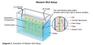
The western blot showed there is a CXCR4 protein being produced in 4T1 cancer cells. The band of protein at 47 kDa throughout the samples run shows there was constantly CXCR4 protein in all of the 4T1 cells tested.
Flow Cytometer
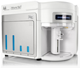
A statistically significant greater number of cells fluoresced with the stain that binds to CXCR4; this implies there is CXCR4 in the 4T1 cells the stain is binding to. This is further evidence there is CXCR4 in 4T1 cells.
Fluorescent Microscope
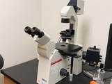
There was a GFP tag attached to the plasmid which glows while under the EGFP light in the microscope. If the cell was glowing under that light, it is probable the plasmid was inserted into the cell. Pictures were taken under a normal and the EGFP light and overlaid to see if there was glowing in the cells.
Cell Sorter
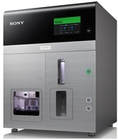
Cells with the plasmid inserted fluoresced more than cells without plasmids did. This sorter then separated out the glowing cells. These cells most likely have the CXCR4 plasmid inserted into them.
Future Studies
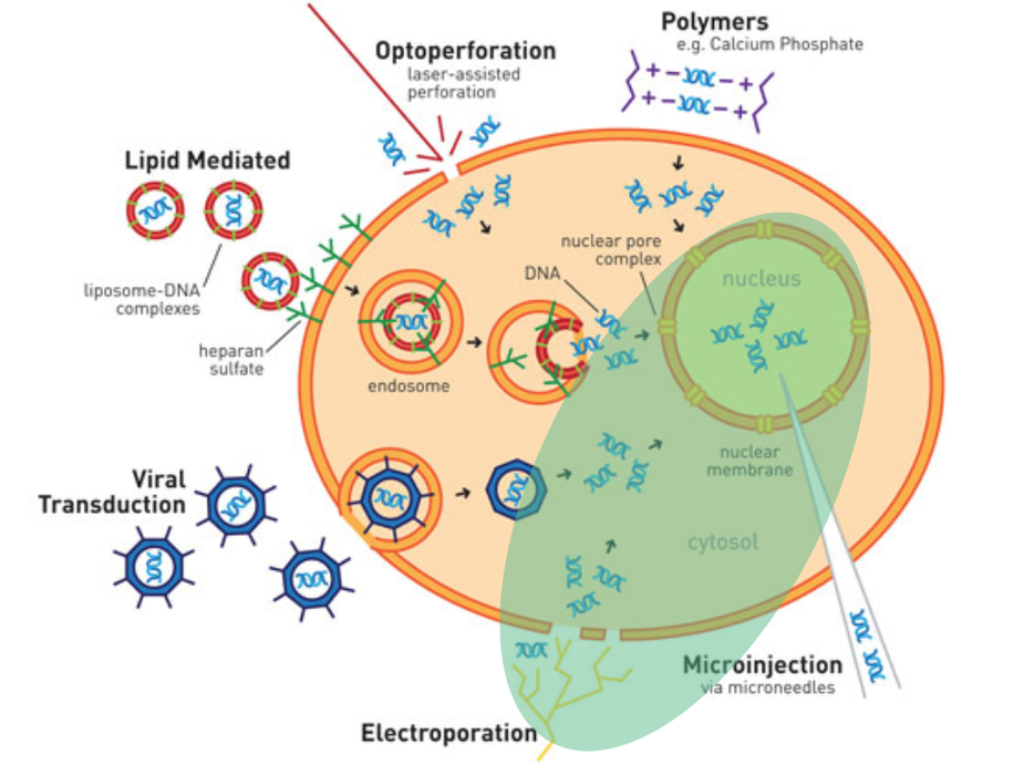
This science project can be continued by testing the cells with the plasmid inserted into them with a western blot to see if the CXCR4 protein is still being produced in the cells. In the future, an electroporation method will be used as a method for transfection. Electroporation is where electricity is run through the cells to create holes in the cell’s membrane so the circular plasmid can go into the cells. This will hopefully have a higher success rate than the previously used lipid mediated transfection method. From there, if transfection is successful and the gene that codes for CXCR4 is successfully knocked down and there are no CXCR4 proteins being produced in the cancer cells, the experiment can then be moved en vivo in murine models.
